Preliminary testing in laboratory animals has long had an essential role in the development of new pharmaceuticals and methods for treating human disease. The current development of sophisticated transgenic animal models as well as a growing recognition of the importance of understanding disease processes in the context of the living host has extended the use of animal experimentation beyond safety and efficacy testing into the realm of mechanistic investigation. Non-invasive imaging makes it possible to perform multiple measurements over time in the same animal, thereby enhancing data quality in studies of dynamic molecular and physiologic processes as well as greatly reducing the number of animals required for such studies.
During the last several years, scanners for small animals have become commercially available for all of the established modalities of medical imaging (X-ray, CT, MRI, SPECT, PET, ultrasound), as well as for optical imaging (Luminescence and Fluorescence). With this technology, the dynamic biodistribution of therapeutic agents as well as vital processes such as gene expression, cell trafficking, cell viability, cell proliferation, tissue hypoxia and angiogenesis can be monitored non-invasively in the intact animal.
Small animal imaging has become indispensable to medical research and development and helps the investigator remain competitive for extramural funding.
Equipment
Imaging systems available are:
The Small Animal Imaging Core is also equipped with a scale and gamma counter (PerkinElmer) which are available for trained users for ex vivo biodistribution studies.
Positron emission tomography (PET) is a nuclear imaging technique that allow visualization, characterization and quantification of biological processes (metabolic and functional) in vivo following administration of a positron emitting radionuclides such as Cu-64, F-18, Ga-68, I-124 or Zr-89. PET can be combined with CT (computed or computerized tomography) that uses X-rays for visualization of anatomical structures (i.e. tissues, organs, and skeletal structure) or MRI (magnetic resonance imaging) that does not use radiation, but a magnetic field and radio waves to create very detailed anatomical structures and superior soft tissue contrast. The PET, CT and MRI images can be viewed separately or as a single, overlapping "fused" PET-CT/MRI image.
Bioluminescence and fluorescence imaging are non-radioactive optical imaging technologies.

https://specimg.com/ https://www.perkinelmer.com/product/ivis-lumina-s5-imaging-system-cls148588
Located in Parvin 1031, Parvin 1037 and BRC 1260
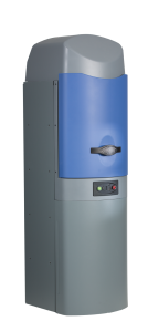
The Lago and Lago X (X-ray capable) and IVIS Lumina S5 are optical imaging systems for both fluorescent and bioluminescent imaging. The light emitted, is captured by a high sensitive cooled charge-coupled device (CCD) camera system of silicon crystal detectors. The free AURA software is used for instrument control and acquisition and analysis.
Bio-luminescence imaging (BLI) results from enzyme-mediated chemical reactions involving injected substrates. The most commonly used enzyme/substrate combination is luciferase/luciferin. Luciferase may be constitutively expressed for monitoring cellular growth and anatomic location, or placed under the control of a promoter of interest and used as a reporter gene. BLI can detect very low levels of signal because the light emitted is virtually background-free.
Fluorescence imaging is created when a molecule (fluorophore, dye) emits light at a certain wavelength after being excited with light at a different wavelength. Both instruments have 14 light-emitting diode (LED) fluorescence excitation filters and 20 emission filters that will cover detection of GFP (509 nm), A488 (519-525nm), mcherry (610 nm), A647 (665 nm), Cy5.5 (695 nm), 800CW (792 nm), ICG (830 nm) and others. More background signal owing to autofluorescence of tissues is seen in fluorescence imaging.
Near-infrared (NIR) fluorescence imaging involves imaging fluorescence photons in the near-infrared range (typically 700–900 nm). NIR fluorophores are used for in vivo imaging because of better photon penetration into tissue, minimal tissue autofluorescence, less scatter, and higher optical contrast (signal-to-background ratios).

https://sofie.com/ https://www.molecubes.com/
Location: Parvin 1037A Location: Hilton 123
GNEXT PET/CT (single gantry)
The GNEXT PET/CT is a high performance and high flexibility preclinical in vivo imaging system that incorporates single or multi-animal (hotel) bed platforms with integrated heating and anesthesia systems. The PET scanner is equipped with dual-layer crystal technology (LYSO and BGO) to overcomes depth-of-interaction (DOI) limitations such as progressive reduction in radial spatial resolution with increasing distance from the center of the FOV that is common in ring PET systems. The PET permits visualization of specific metabolic and physiological processes with high sensitivity (>12%) and spatial resolution (<1mm) at the center of the field of view (FOV). The CT camera has 3072 x 1944 pixels and a magnification range of 1.3 – 5.0. The focus X-ray source has a maximum resolution of 20µm. The standard scan time is ~1 minute. The system has a bore size of 13.9 cm and an axial FOV of 10.4 cm that can be extended to 30 cm. All PET and CT data is collected in list mode format and images are stored in DICOM format that can be exported and analyzed in any imaging software such as VivoQuant and AMIDE. The scanner will acquire and reconstruct your data simultaneously using OSEM reconstruction protocols.
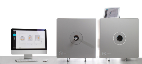
MOLECUBES PET/CT (modular system)
The MOLECUBES is a high performance and high flexibility modular benchtop preclinical imaging system that also incorporates single or multi-animal (hotel) bed platforms with integrated heating and anesthesia systems. The system comes with an integrated preparation stage that is coupled to the anesthetic flow and can be put next to the scanner. This stage facilitates the preparation of both rats and mice, including the insertion of multiple catheter lines and administration of tail vein and subcutaneous injections.
The PET imaging module provides sub-millimeter image resolution (0.85mm) through the combination of monolithic scintillators, the latest photon counting technology and GPU-based event positioning (LYSO crystals with SiPM technology), and iterative image reconstruction (OSEM). The 5-ring configuration ensures best-in-class sensitivity (>12%) over a field of view (FOV) that is adequate for whole-body mouse and rat imaging at a high count rate. In-house hardware enables dynamic and respiratory and/or cardiac gated studies with temperature monitoring. All PET data is collected in list mode format and images are stored in DICOM format that can be exported and analyzed in any imaging software such as VivoQuant and AMIDE.
The CT imaging module allows for fast whole body mouse and rat CT imaging at extremely low dose (<4 mGy) and excellent soft tissue contrast. Advanced workflows, such as gated and dynamic contrast enhanced imaging, can be achieved in a functional and integrated setup. Iterative reconstruction techniques are available in standard as well as in expert user mode. Maximum resolution is 50µm.
This modular system facilitates synchronous scanning. Since the modalities are separated into different units, one modality does not block another during the scanning process, as is the case with single gantry systems.
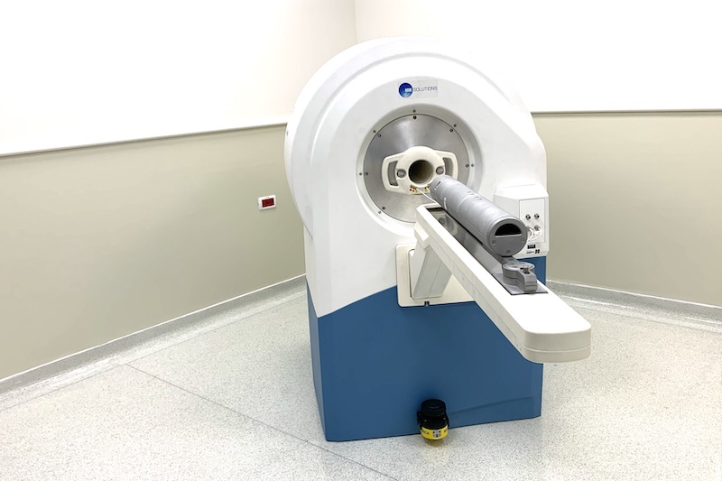
7TMRI/PET (MRSolutions)
Location: Parvin 1037A
7T simultaneous PET-MRI is a cryogen-free, dual modality imaging system specifically designed for in vivo and/or ex vivo applications. The magnet in the 7T Benchtop MRI system has variable field strength (0.1T – 7.0T) for superior soft tissue contrast, and provides high spatial resolution for great visualization and quantification. The scanner can be configured with high gradient strength, and advanced coils such as multinuclear, phased array etc. The simultaneous acquisition of PET and MR images combines the high sensitivity of PET with the high spatial resolution and soft tissue contrast of MRI. For simultaneous PET and MR imaging, the PET module is inserted within the bore of the MRI system for automatic imaging from one modality to another on the same axis. The PET is equipped with dual-layer LYSO crystals that provides image resolution at <0.8 mm at the center of FOV. The 7TMRI/PET bore size is 24 cm and the PET axial FOV is 10.04 cm. Images are exported in DICOM format and reconstructed with 3D-OSEM format that can be analyzed in any imaging software such as VivoQuant and AMIDE. The system is provided with Powerscan software permitting access to all functions of the system.
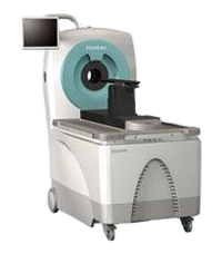
Location: Gonda B1016
The Inveon PET imaging system has 10 mm thick LSO crystals with 1.59 mm x 1.59 mm detector pixel spacing resulting in an absolute sensitivity of 10% at the center of the field of view. The system has an axial field of view (FOV) of 12.7 cm that is extended to 30 cm with continuous bed motion. The system is provided with Inveon Acquisition Workplace (IAW) and Inveon Research Workplace (IRW) software for image acquisition and processing, 2/3-D image viewing, and quantitative analysis. A variety of image reconstruction algorithms are provided (2D FBP, 3DRP, OSEM2D, OSEM3D/MAP) format that can also be analyzed in any imaging software such as VivoQuant and AMIDE.
As an NCI-designated Comprehensive Cancer Center supported Core facility, it is critically important to acknowledge the Core Cancer Center Support Grant (CCSG) if you’ve used any of our imaging instruments and/or services for publications, presentations, grant applications, media releases, or any other form of media.
As a courtesy, we also request that you inform us of the publication.
"Research reported in this publication included work performed in the Small Animal Imaging Core supported by the National Cancer Institute of the National Institutes of Health under grant number P30CA033572. The content is solely the responsibility of the authors and does not necessarily represent the official views of the National Institutes of Health."
The Small Animal Imaging Core (SAIC) is directed by Dr. Anna Wu, managed by Dr. Tove Olafsen and staffed full-time by Leslie Rodriguez and part-time by Dr. Weidong Hu.
| Hours | Location |
|
8:30 am - 5:00 pm Monday - Friday
6:00 am - 6:00 pm for Lago, Lago X and IVIS after Training |
Parvin 1031, Parvin 1037, Parvin 1037A, BRC 1260 and Hilton 123 |